
Fig. 1.
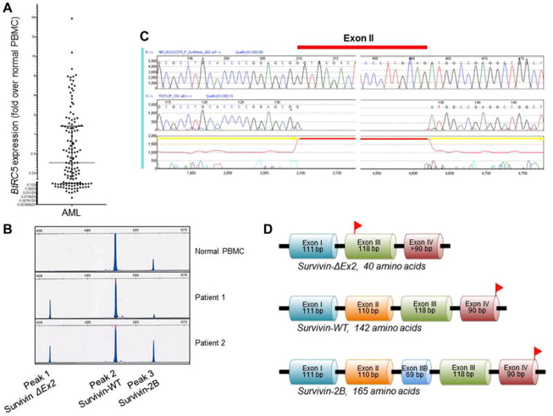
Identification of survivin-2B and the novel splice variant ΔEx2. A: Range of BIRC5 expression overall, relative to normal PBMCs. B: Capillary electropherogram from normal PBMCs and two representative patients with 2B/ΔEx2 ratios <1 (Patient 1) and >1 (Patient 2). C: Sequence analysis revealed loss of exon II from the splice variant represented by peak 1 in the electropherogram (B), and the variant was hence termed survivin-ΔEx2. D: Deletion of exon II results in a frameshift mutation, leading to a premature stop codon in exon III. Stop codons are represented by red flags. PBMCs, peripheral blood mononuclear cells; WT, wild-type; bp, base pairs.