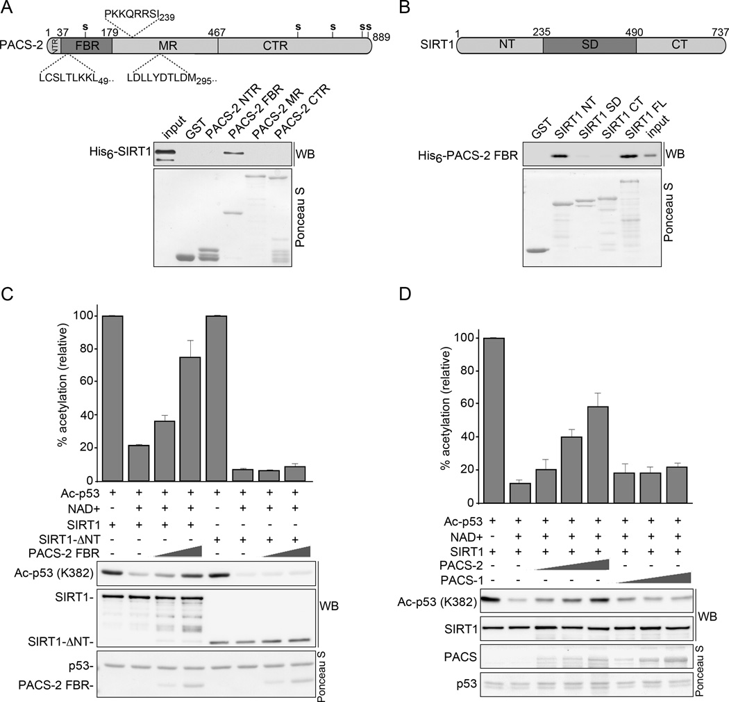
Figure 7. PACS-2 inhibits SIRT1-mediated deacetylation of p53 in vitro.
(A) Top: Schematic of hPACS-2 and the predicted NLS (above) and NES (below). Bottom: Full length His6-SIRT1Δ6–83 was incubated with GST or GST-PACS-2 constructs and interacting His6-SIRT1Δ6–83 was detected by western blot. Captured GST fusion proteins detected with Ponceau S. (B) Top: Schematic of mSIRT1. Bottom: His6-PACS-2FBR was incubated with GST or GST-SIRT1 constructs and interacting His6-PACS-2FBR was detected by western blot as in (A). (C) GST-Ac-p53 (see Methods) was incubated with 0.5 µM NAD+ and His6-tagged SIRT1 or SIRT1-ΔNT together with increasing amounts of His6-PACS-2FBR as indicated. The amount of Ac-p53 (K382) was quantified and presented as percent acetylation. Error bars represent mean ± SEM from 4 independent experiments. (D) GST- Ac-p53 was incubated with 0.5 µM NAD+ and His6-SIRT1 together with increasing amounts of His6-PACS-1 or PACS-2 as indicated. The amount of Ac-p53 (K382) was quantified as in (C). See also Figure S4.

