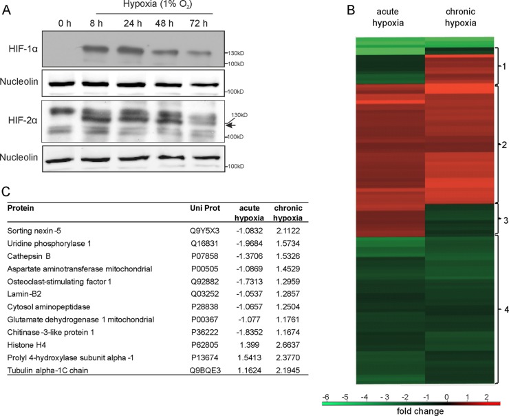
FIG 1.
Protein expression in response to acute versus chronic hypoxia. (A) Western analysis of HIF-1α and HIF-2α in primary human macrophages incubated for up to 72 h under hypoxia. Expression of nucleolin serves as a loading control. (B) Heat map showing protein expression of 2D-DIGE experiments performed with THP-1 macrophages incubated in acute (8 h) or chronic (72 h) hypoxia. (C) List of the 12 proteins identified by MS/MS from cluster 1 of panel B that are expressed under chronic hypoxia. Expression values are ratios between control conditions and acute or chronic hypoxia.

