Figure 2.
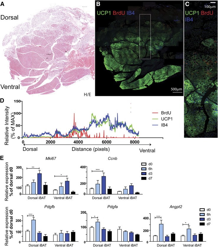
Cold stress triggers cell proliferation at the dorsal edge of BAT that interfaces suprascapular WAT. A and B) Histologic analysis of BrdU incorporation in iBAT during cold exposure. Adjacent paraffin sections were stained for H/E (A), or BrdU, isolectin-IB4, and UCP1 (B). Scale bar, 500 µm. C) Magnified view of boxed region from (B). Scale bar, 100 µm. D) Analysis of fluorescence intensity of (C) indicated that cell proliferation (BrdU-Red) concentrated at the dorsal edge of BAT, where UCP1 expression and vascular density drop. MAX, maximum. E) Quantitative PCR analysis of proliferation- and angiogenesis-related gene expression in dorsal and ventral BAT of control mice and mice exposed to 4°C up to 7 d (n = 6, mean ± sem). *P < 0.05; **P < 0.01;***P < 0.001.

