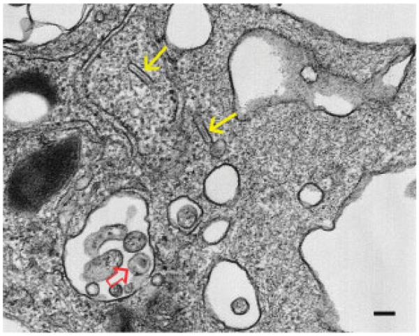
Figure 1.
Transmission electron microscopy of a human LC, as identified by the rod-shaped Birbeck granules (yellow arrows), that was exposed to HIV-1 in vitro. The cell contains one clearly discernible HIV-1 particle (open red arrow). This is the “stage” where the article by Geijtenbeek and colleagues “plays.” Diameter of the HIV-1 particle and the scale bar is about 100 nm.

