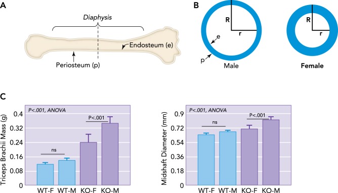
FIGURE 1.
Schematic representation of a long bone and dimorphism in bone cross-sectional geometry
A: schematic representation of a long bone indicating the outer periosteal surface (p) and inner endocortical surface lining the medullary cavity (e), with a dotted line showing cross section. B: schematic representations of long bone cross-sectional geometry in males vs. females at the level of the dotted line shown in A. Males typically have a wider bone cross section and outer radius (R), whereas females have a smaller outer radius but thicker bone cortex, leading to a shorter inner radius (r). As women age, bone is resorbed from the endocortical surface so that older women must increase R to compensate for an increasing r. Males have a larger R and so are at lower fracture risk even as r increases. C: increased muscle mass in males is associated with increased bone diameter, even in mice. Male mice (M) lacking myostatin show a greater increase in forelimb triceps brachii mass than female (F) knockout (KO) mice, and forelimb bone (radius) diameter is largest in M mice lacking myostatin (KO) compared with wild-type (WT) mice.

