
FIGURE 1.
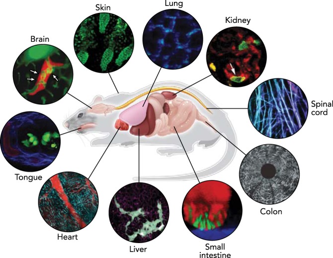
In vivo imaging in the mouse
Advances in optical techniques in conjunction with sample preparation methods allow microscopic visualization of cellular dynamics in various organs in a living experimental animal. Representative images in each organ acquired by intravital microscopy on mouse models are shown. Images reproduced with permission from Refs. 55 (brain), 92 (ear skin), 75 (lung), 23 (kidney), 29 (spinal cord), 61 (colon), 89 (small intestine), 90 (liver), 50 (heart), and unpublished observations of Choi M, Lee WM, Yun SH (tongue).