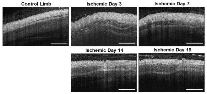
Fig. 5.

Representative structural OCT images from the adductor muscle region, including the contralateral control limb (left) and the ischemic limb imaged non-invasively over a time-course. A change in tissue structure beneath the skin is observed in the ischemic limb, presumably due to inflammation. Scale bar is 1 mm.
