
Fig. 1.
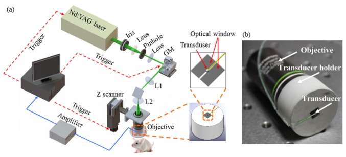
(a) Schematic diagram of the reflection-mode subwavelength-resolution photoacoustic microscopy system. (b) Photograph of the configured imaging probe consisting of a high-NA objective and a miniature high-frequency ultrasonic transducer. GM: galvanometer scanner; L1: scan lens; L2: tube lens.