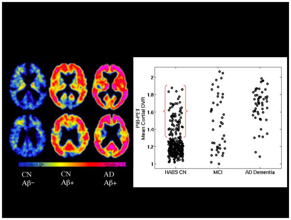
Figure 1.
PET amyloid imaging with 11C-PiB. Left: Representative PET images from three older individuals: Clinically normal older individual without evidence of elevated Aβ accumulation (CN Aβ−), Clinically normal older individual with elevated Aβ accumulation (CN Aβ+) and patient with AD dementia with very elevated Aβ accumulation (AD Aβ+) in frontal and parietal heteromodal cortices Right: Scattergram of PiB distribution value ratios (DVR) by diagnostic group: Harvard Aging Brain Study Clinically normal older individuals (HABS CN), Mild Cognitive Impairment (MCI), and AD dementia. Approximately 30% of HABS CN demonstrate elevated Aβ accumulation in the range of MCI and AD dementia Aβ+.

