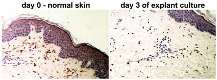
Fig. 3.
Human skin before (left) and after a 3-day skin explant culture (right). Dermal macrophages were visualized with an immunoperoxidase technique using antibodies against Factor XIIIa, a marker for these cells (Zaba et al. 2007). Positive cells can be identified by the brown reaction product (few examples marked with an asterisk). Note that after 3 days of culture the numbers of dermal macrophages are markedly reduced

