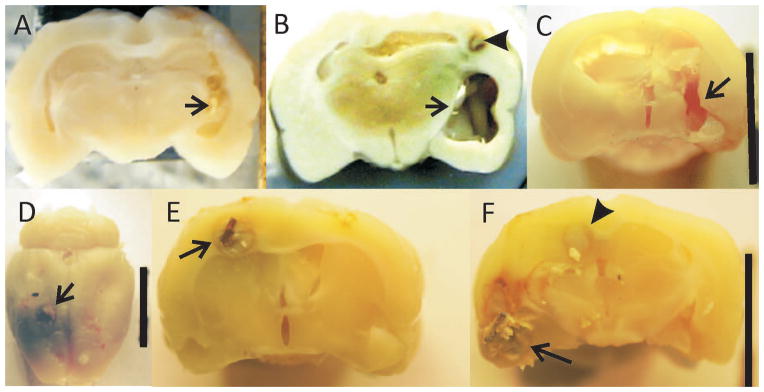
Figure 5.

Gross sections showing the lesions from 3 rats. Arrows show lesion. A. Lesion only animal without epilepsy. Lesion consists of the drill tract with little addtional damage. B. Lesion plus copper after 7 months in rat with documented epilespy. There is significant tissue loss in the medial-lateral as well as anterior posterior directions extending far beyond the primary lesion. In addition, precipitated copper discolors the surrounding tissue. Arrowhead points to copper wire remnant surrounded by necrotic tissue. C. Lesion only animal with epilepsy. Lesion more extensive than in animal in (A) extending to the ventral surface as well as anterior and posterior to primary lesion in (A). D–F: Brain from an unmonitored rat with lesion plus copper two months after lesion. D. Arrow points to copper wire at cortical surface surrounded by precipitated copper. E. Copper wire surrounded by necrotic tissue. F. Same animal approximately 3mm posterior to (E) More extensive necrosis in region of amygdala and olfactory cortex (arrow) and area of necrosis (arrowhead) continuous with same area in (E). D–F demonstrate the early stages of the copper associated damage.
