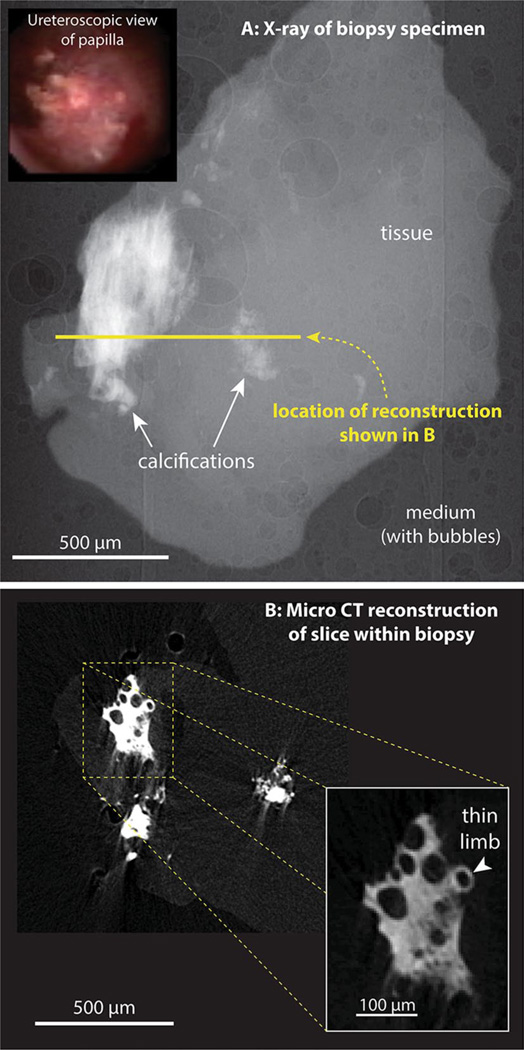
Fig. 1.
Micro-CT imaging of Randall’s plaque in human papillary biopsy. a Inset ureteroscopic image of papillary surface before biopsy. a X-ray image of biopsy. About 450 such images were taken, with the specimen rotated 0.4° between each image. This image series was used for tomographic reconstruction. b Reconstructed slice through the biopsy, with inset showing higher magnification of a small portion of Randall’s plaque within the tissue

