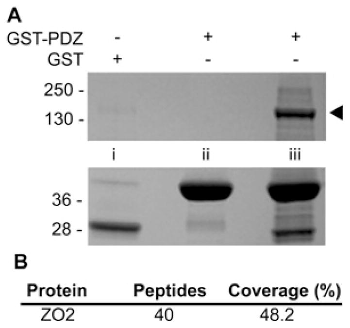
Figure 1. Identification of ZO-2 as a potential SNX27 PDZ binding partner.

(A) mpkCCD cell extracts (lanes i and iii) or lysis buffer (lane ii) were incubated with GST–SNX27 PDZ (lanes ii and iii) or GST controls (lane i). Bound proteins were separated by SDS/PAGE and identified by Coomassie Blue staining. Prominent bands between 28 and 40 kDa are GST fusions. Band marked by arrow was excised, in-gel digested with trypsin and identified as ZO-2 by MS. Molecular mass is shown on the left-hand side in kDa. (B) ZO-2 peptide coverage statistics.
