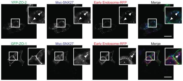
Figure 3. Recruitment of ZO-2, but not ZO-1, to the early endosome by SNX27.

mpkCCD cells were infected with RFP–Rab5a (early endosome marker) and co-transfected with Myc–SNX27 and either YFP–ZO-2 (upper panels) or YFP–ZO-1 (lower panels) infected with RFP–Rab5a and grown to confluence. At this point, the cells were fixed/permeabilized and stained with an anti-Myc monoclonal antibody and visualized using Alexa Fluor® anti-rat 647 nm. Arrows indicate co-localized protein (scale bar = 15 μm).
