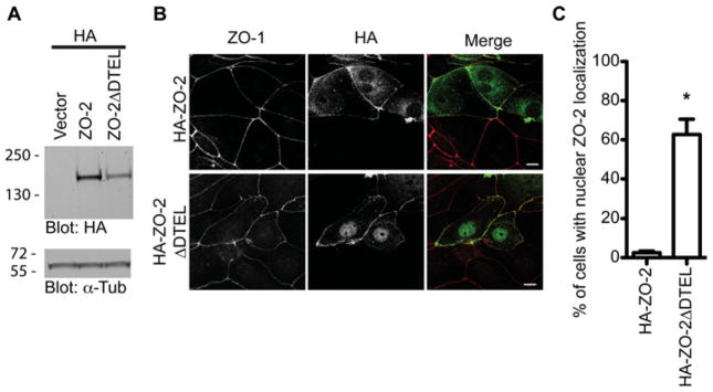
Figure 6. C-terminal PDZ-binding motif of ZO-2 is important for localization.

(A) Western blot of indicated HA-tagged ZO-2 constructs in mpkCCD cells. Equal loading was confirmed by Western blotting using an anti-α-tubulin antibody. (B) Localization of HA–ZO-2 fusion proteins was assessed in mpkCCD cells by staining with an anti-HA monoclonal antibody and anti-ZO-1 polyclonal antibody (scale bar = 10 μm). (C) The proportion of cells expressing nuclear HA–ZO-2 in mpkCCD cells was quantified. The histogram is representative of three independent experiments (Student’s t test, *P < 0.005, n>100; error bars represent the S.D.).
