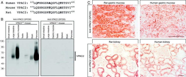
Figure 2.
Characterisation of SP235 using mouse, rat and human tissues. (A) Comparision of the carboxyl-terminal sequences of mouse, rat and human VPAC2. The sequence depicted for the human VPAC2 was used for antibody generation. (B) Western blotting analysis of SP235 in various tissues. Tissue extracts from WT mice (Vpac+/+) and mice lacking Vpac2 (Vpac2−/−) were separated on 7.5% SDS–polyacrylamide gels and blotted onto PVDF membranes. Membranes were then incubated with the rabbit monoclonal anti-VPAC2 antibody (SP235) at a dilution of 1:500. Membranes were developed using ECL. Ordinate, migration of protein molecular weight markers (kDa). (C) SP235 immunohistochemistry in rat and human tissues. Sections were dewaxed, microwaved in citric acid and incubated with the rabbit monoclonal anti-VPAC2 antibody (SP235) at a dilution of 1:500. Sections were then sequentially treated with biotinylated anti-rabbit IgG and AB solution. Sections were then developed in AEC and lightly counterstained with haematoxylin. Representative photomicrographs from one of five different tissue samples are shown. Scale bar: upper panel=200 μm and lower panel=100 μm.

 This work is licensed under a