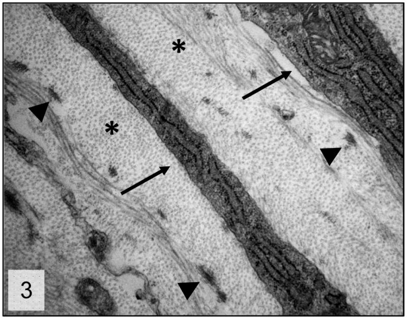
Figure 3.

Trabecular meshwork; 2 week-old wild-type mice. Higher magnification presenting the normal relationship between the trabecular beam’s components. Collagen fibers (*), trabecular cells cytoplasmic projections (arrows) and elastic fiber aggregates (arrow heads). Transmission electron microscopy, uranyl acetate. 8800x.
