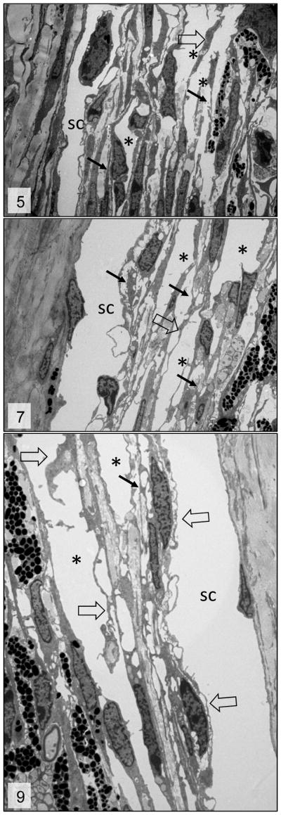
Figures 5, 7 and 9.

Trabecular meshwork; 3 week, 6week and 3 month-old Cyp1b1−/− mice, respectively. Irregular trabecular beams with accentuation of the inter-trabecular spaces (*), fragmentation of the collagen fibers (arrows) and irregular and reactive trabecular cells (open arrows). sc = Schlemm’s canal. Transmission electron microscopy, uranyl acetate. 2650x.
