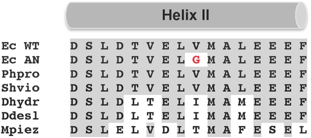
Figure 6.

Multiple sequence alignment of helix II region of various ACP. Identical residues are shaded gray, while red is used to highlight the glycine in AN62 strain. Ec WT, E. coli parental strain; Ec AN, E. coli AN62 strain; Phpro, Photobacterium profundum SS9; Shvio, Shewanella violacea DSS12; Dhydr, Desulfovibrio hydrothermalis AM13; Ddesl, Desulfovibrio desulfuricans 27774; Mpiez, Marinitoga piezophila KA3.
