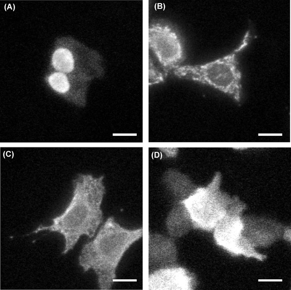
Fig 5.

Bioluminescence images of NanoLuc fused with NLS (A), CoxVIII (B), calreticulin (C) or no targeting sequence (D) in U2OS cells. Images were captured using an LV200 microscope with UPlanFLN 100× Oil objective lens and ImagEM EM-CCD camera at 37°C. Exposure time, 300 ms (A, D), 500 ms (B) and 1 sec (C); Furimazine, 12.5 μM; Scale bars, 20 μm.
