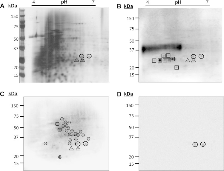
FIG 2.
Identification of the langerin-reactive M. leprae cell wall glycoprotein SodC. The cell wall protein fraction of M. leprae (MLCwA) was resolved by 2D PAGE and silver stained (A) or transferred to PVDF membranes and probed with ConA (B), r-langerin (C), or rabbit anti-SodC polyclonal sera (D). The image for the ConA blot was obtained from 20 s of exposure time and the r-langerin blot from 5 min of exposure time in the ChemiDoc XRS system. Circles and triangles indicate the 2D PAGE locations of M. leprae SodC and SodA, respectively. Hexagons designate the other protein spots reactive to r-langerin, and squares designate ConA reactive spots.

