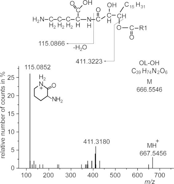
FIG 6.

Q-TOF MS/MS spectrum of OL-OH analyzed in the positive-ion mode. The fragment pattern represents a main molecular OL-OH species with a 16:0 fatty acid esterified to the primary hydroxy group of an amide-bound dihydroxy-18:1 fatty acid. The exact position of the secondary hydroxy group cannot be determined by Q-TOF MS/MS. The protonated form of this species (parental ion) yields a mass of m/z 667.5456 (calculated mass is 667.5618 u). The neutral loss of 256.2276 u (667.5456 u minus 411.3180 u) is derived from 16:0 as an ester-linked fatty acid. The fragment ion m/z 411.3180 represents the lyso form of this ornithine lipid containing the additional hydroxy group linked to the amide-bound 18:1-hydroxy fatty acid. The low-mass fragment ion at m/z 115.0852 is consistent with the presence of ornithine in this lipid (45), forming a ring structure with loss of a water molecule (COO-R1 represents a 16:0 acyl chain).
