
Fig 4.
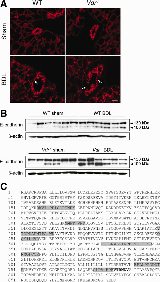
E-cadherin expression in Vdr−/− mice. (A) Representative immunostaining of E-cadherin in the liver of Sham or BDL, wildtype (WT), and Vdr−/− mice 3 days post-BDL. E-cadherin staining was interrupted (arrows) more often in BDL Vdr−/− (lower right panel) than in WT mice (lower left panel), indicative of higher incidence of bile duct rupture (33.0 ± 0.04% and 14.8 ± 0.08% of bile ducts, respectively). Scale bar = 10 μm. (B) Liver samples were collected from Sham or BDL, WT, and Vdr−/− mice 3 days post-BDL and were assessed for E-cadherin expression by immunoblot. β-Actin was used as an internal control. (C) Whole liver protein extracts were immunoprecipitated with an antibody directed against E-cadherin. After gel migration, the 100-kDa protein signal was extracted and submitted to mass spectrometry analysis. The full E-cadherin protein sequence is shown, with dark boxes representing the peptides identified by mass spectrometry. The underlined sequence corresponds to the calpain cleavage site consensus sequence.