Figure 1.

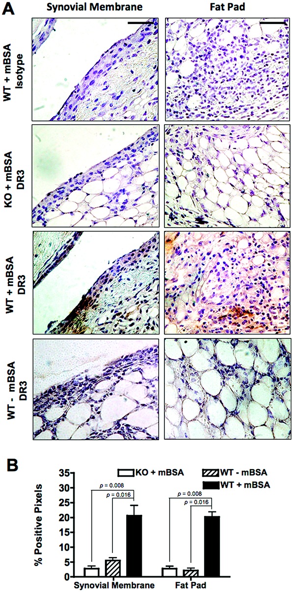
Death receptor 3 (DR-3) expression in the joints of mice with antigen-induced arthritis. Arthritis was induced in DR3-knockout (DR3-KO; DR3−/−) mice and their DR3+/+ (wild-type [WT]) littermates, and the joints were prepared, sectioned, and stained for DR-3 as described in Materials and Methods. Antigen (methylated bovine serum albumin [mBSA]) was administered into the right knee to induce localized inflammatory arthritis. A, Representative high-magnification (40×) photomicrographs showing (from top to bottom) isotype staining in a right knee section from a WT mouse, anti– DR-3 staining in a right knee section from a DR3−/− mouse, anti–DR-3 staining in a right knee section from a WT mouse, and anti–DR-3 staining in a left knee section (contralateral negative control) from a WT mouse. Bars = 45 μm. B, Quantification of positive staining, as measured by the percentage of positive pixels within a particular area. Values are the mean ± SEM (n = 4–5 mice per group). P values were determined by Mann-Whitney U test.
