
Figure 2.
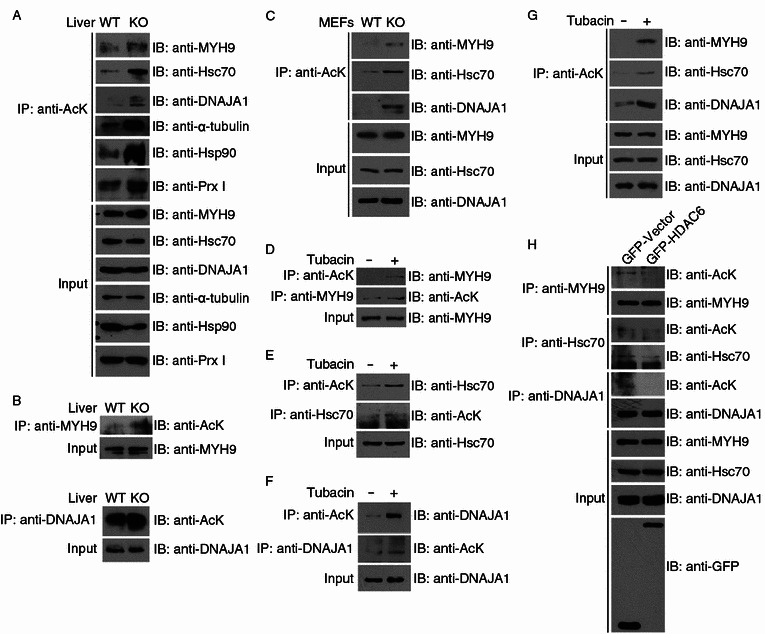
HDAC6 deacetylates selected substrate candidates. (A) Soluble cytoplasmic proteins were extracted from the liver tissues of wild-type and HDAC6 knockout mice, and immunoprecipitation was performed with anti-AcK antibodies. The immunoprecipitates and tissue extracts were then immunoblotted with the indicated antibodies. (B) Protein samples were prepared as in (A), and immunoprecipitation was performed with anti-MYH9 or anti-DNAJA1 antibodies. The immunoprecipitates and tissue extracts were then immunoblotted with the indicated antibodies. (C) Total cell lysates were prepared from wild-type and HDAC6 knockout MEFs. Anti-AcK immunoprecipitates and cell lysates were immunoblotted with antibodies against MYH9, Hsc70, or DNAJA1. (D–F) 293T cells were treated with DMSO (−) or tubacin (+) for 8 h. Anti-AcK, anti-MYH9, anti-Hsc70, or anti-DNAJA1 immunoprecipitates and total cell lysates were immunoblotted with the indicated antibodies. (G) Cytoplasmic proteins were extracted from 293T cells treated with DMSO (−) or tubacin (+) for 8 h. Anti-AcK immunoprecipitates and cytoplasmic extracts were immunoblotted with antibodies against MYH9, Hsc70, or DNAJA1. (H) 293T cells were transfected with GFP-HDAC6 or GFP alone. Immunoprecipitation and immunoblotting were then performed with the indicated antibodies