
Figure 3.
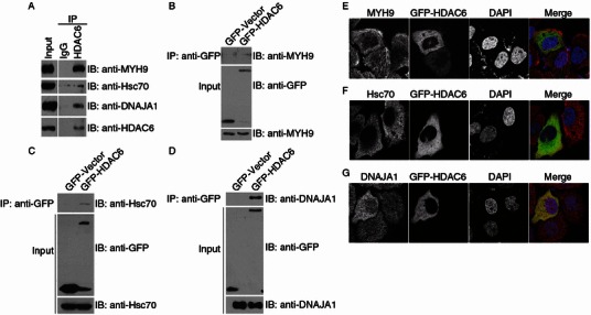
HDAC6 interacts with the new substrates. (A) 293T cell lysates were incubated with control IgG or anti-HDAC6 antibodies. The immunoprecipitates and cell lysates were then immunoblotted with the indicated antibodies. (B–D) 293T cells were transfected with GFP-HDAC6 or GFP alone. Anti-GFP immunoprecipitates and cell lysates were immunoblotted with the indicated antibodies. (E–G) Immunofluorescence confocal images of HeLa cells transfected with GFP-HDAC6 and stained with anti-MYH9 (E), anti-Hsc70 (F), or anti-DNAJA1 (G) antibodies and the DNA dye DAPI