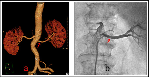
Figure 3.

(a) Multidetector computed tomography angiography of bilateral renal arteries and 3‐dimensional imaging of renal artery. The arrow points to the middle section of the right renal artery where atherosclerosis plaque could be observed. The stenosis of the renal artery was 32.8% and plaque was present. (b) The right renal arteriography during radiofrequency renal denervation of the same patient. The arrow points to the middle section of the right renal artery where atherosclerosis plaque could be observed.
