
Figure 2.
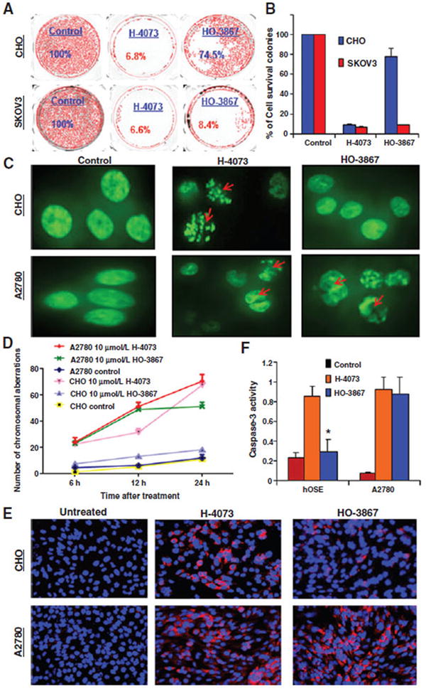
Selective cytotoxicity of HO-3867 toward to cancer cells. A, clonogenic assay: SKOV3 and CHO cells were treated with 10 μmol/L of H-4073 or HO-3867 for 24 hours, after which the drug was removed and cells were observed for 72 hours for colony-forming ability. B, quantification of colony survival. C, A2780 and CHO cells engineered to express a GFP-Histone 2B fusion protein were treated with 10 μmol/L of H-4073 or HO-3867 at different time points, followed by immunofluorescence microscopy. Red arrows, individual cells that underwent abortive mitosis—with transient chromatin condensation or apoptosis. D, quantification of chromosomal aberration at 6, 12, and 24 hours following treatment reveals that H-4073 causes more chromosomal aberration than HO-3867 in CHO cells (P < 0.0001 at all-time points) but not in A2780 cells where a difference only developed at 24 hours (P = 0.9982, 0.9216, <0.0001). E, immunofluorescent staining using 8-hydroxyguanosine (8OHdG), after treating cells with HO-3867 or H-4073 for 6 hours. Before treatment neither CHO nor A2780 stained positive for 8OHdG. After treatment with H-4073, both cell lines showed similarity to 8OHdG. Conversely, HO-3867 treatment induced elevated 8OHdG in A2780 relative to CHO cells (red, 8OHdG; blue, DAPI nuclear stain). Magnification, ×200. F, HO-3867 induced more caspase-3 activity in A2780 than hOSE cells compared with H-4073 (*, P ≤ 0.0001, n = 3).