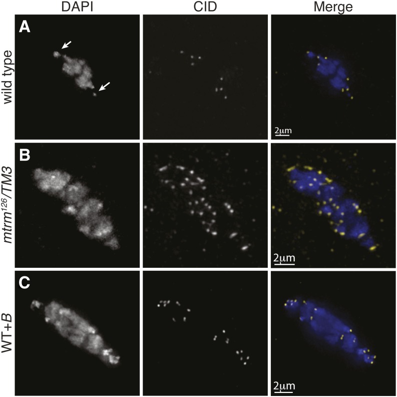
Figure 1.
B chromosomes contain centromeres. Fixed prometaphase I oocytes were prepared with antibodies against CID (yellow) and the DNA dye DAPI (blue). (A) Wild-type oocytes display the expected 8 CID foci. Arrows indicate the location of the 4th chromosomes. (B) A mtrm126/TM3 oocyte displaying 37 CID foci (mean of 29.3, SD 7.5, range 10–43, n = 49). (C) A WT+B oocyte with 19 CID foci.

