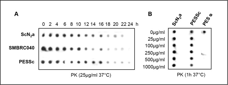
Figure 2. Relative resistance of PESSc prions to ProteinaseK.

A) Cell lysates from PESSc cells, ScN2a cells and SMBRC040 cells were each divided into 13 aliquots. All aliquots, but one of each cell line, were subjected to ProteinaseK digestion (25 µg/ml; at 37°C). Every two hours the digestion of one aliquot per cell line was stopped with PMSF. PrPres was detected by dot-blot analysis (mAB ICSM18). In both murine cell lysates PrPres was detectable for up to 22 hours; in PESSc cell lysates PrPres was still detectable after 24 hours. B) Cell lysates from uninfected PES cells, PESSc cells and ScN2a cells were divided into six aliquots each, ProteinaseK was added at increasing concentrations (0 µg/ml, 25 µg/ml, 100 µg/ml, 250 µg/ml, 500 µg/ml and 1000 µg/ml), and the samples were incubated for one hour at 37°C. The digestion was terminated by adding PMSF. PrPres was detected by dot-blot analysis (mAB ICSM18). While 25 µg ProteinaseK/ml were sufficient to clear the control lysate from any detectable PrP, strong PrPres signals were detected in both prion infected samples up to 500 & 1000 µg ProteinaseK/ml.
