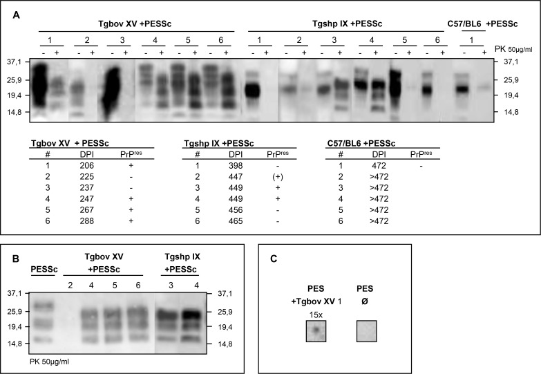
Figure 4. Verification of infectivity in PES cells by mouse bioassay.
A) Cell lysates of PESSc cells were inoculated into three groups of mice (six animals each group): Tgbov XV, Tgshp IX and C57/BL6. Tgbov XV mice succumbed to disease 245 ± 29 dpi, Tgshp IX mice died 444 ± 23 dpi and the first non-transgenic mouse was culled due to clinical signs 472 days post inoculation. The brain homogenates were tested by western-blot analysis for PrPres signals. B) Side by side western-blot analysis of PK digested PrPres derived from PESSc cells shows a higher molecular weight than PK digested PrPres from PESSc infected Tgbov XV or Tgshp IX mice. C) PES cells were inoculated with brain homogenate from Tgbov XV mouse 1. The cells were split 15 times at a ratio of 1:2 and were then assayed for PrPres by dot-blot analysis. Uninfected PES cells were assayed as control Ø. mAB: ICSM18.

