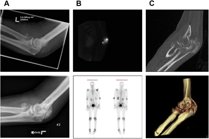
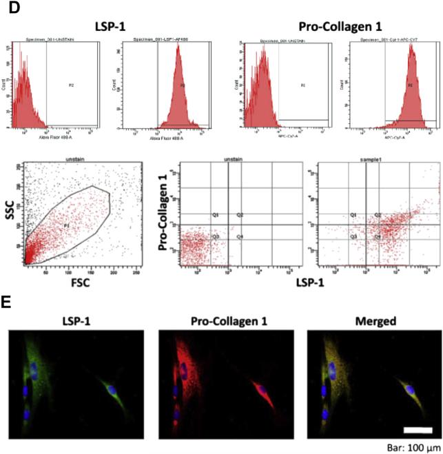
Fig. 2.
A 26-year-old man with 75% TBSA burns who developed HO in both elbows. (A) Imaging studies of the right elbow at 1.5 months (left) and 5 months (right) after burn injury demonstrate the progression of the HO lesion. (B) Intraoperative views from the same patient showing the surgical approach for HO resection (left) and HO specimen (right). (C) Isolation of bone marrow–derived precursor cells from HO tissue by using cell explantation method. A significant cell subset isolated from HO tissue (~35–65%) exhibits a LSP11/COL11 profile as demonstrated by flow cytometry (D) and immune fluorescence microscopy (E).


