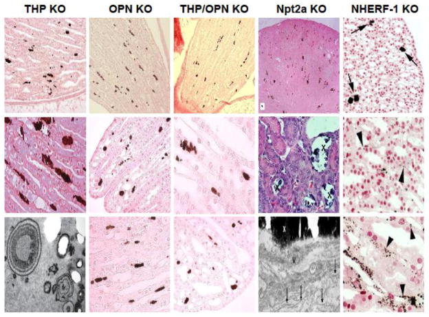
Figure 1.
Morphological features of renal papillary interstitial calcinosis in knockout (KO) mice lacking Tamm-Horsfall protein (THP), osteopontin (OPN), THP and OPN, Na+-phosphate cotransporter Type II (Npt2a) and Na+/H+ exchanger regulatory factor (NHERF-1). Histochemistry for the detection of calcium crystals was von Kossa method for all the KO mice except for NHERF-1 where Yasue staining was used. The lower panels also contained transmission electron micrographs for THP KO and Npt2a mice. IX and X in the middle and lower panels of Npt2a KO mice denote interstitial crystals. The images were adapted, reoriented and resized from the original publications with permission (THP KO from (ref. 62); OPN KO from (ref. 62); THP/OPN KO from (ref. 62); Npt2a KO from (ref. 105); and NHERF-1 from (ref. 65)).

