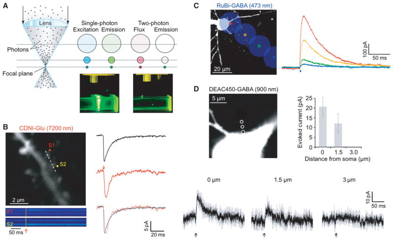
Fig. 4.
Introduction to 1P and 2P uncaging. (A) Left, cartoon of a lens focusing light. Middle, 1P excitation occurs throughout the light absorption path and total excitation is equal in each z-section. Right, 2P emission is confined to the focal plane due to the nonlinear nature of the creation of the excited singlet state. (B) Left, image of a dendritic segment of a CA1 pyramidal cell filled with Oregon Green BAPTA-1 (0.2 mM) using 2PE at 820 nm showing spines that were targeted for 2P uncaging of CDNI-Glu (1 mM) with 720 nm light (50 mW). Spine-selective 2P uncaging of CDNI-Glu was revealed by line-scan imaging (dotted line) with a period of 0.7 ms (146 pixels, 4 μs/pixel). Increases in [Ca2+] are shown in pseudocolor traces and reveal that S1 and S2 were stimulated separately. Right, currents (red traces, n = 24 from five cells) from uncaging of CDNI-Glu (1 mM, 720 nm, 1 ms) at single spines closely mimicked miniature EPSCs (black traces, n = 108). (C) A 473-nm laser was targeted on a cell soma and, at 20-μm increments (colored stars), the corresponding current traces from photolysis of RuBi-GABA (10 μM, 2 ms, 20 mW) are shown. (D) Two-photon photolysis of DEAC450-GABA (0.2 mM, 900 nm, 5 ms, 100 mW) evoked outward currents that decreased as the laser was moved away from the cell body. Each trace is an average of three events recorded from three cells, with grey indicating the SEM. For C and D, CA1 pyramidal cells were held at −40 mV. Compared to 1P uncaging, 2P uncaging generates a much smaller spatially restricted input. Brian slices were isolated as described in the Appendix.

