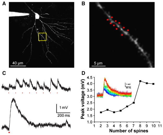
Fig. 5.
Temporal integration of excitation at multiple spine heads. (A) Two-photon fluorescence image of a CA1 pyramidal cell. (B) Detail of dendrite shown in the yellow box in (A), with a cluster of 10 spines targeted for the experiment in (D). (C) CDNI-Glu (1 mM) was uncaged at 720 nm at eight spines along a dendrite, compressing the inter-pulse interval between uncaging events from 100 ms (upper) to 1 ms (lower), producing a nonlinear response. Individual responses from each spine varied between 0.25 and 1.0 mV, and temporal clustering doubled the integrated voltage response. A similar nonlinear dendritic response (dendritic spike) can be clearly seen in (D), where the inter-pulse interval was 0.12 ms. (D) Plot of the somatic voltage responses from uncaging at two to ten spines, with the associated voltage traces inset. The evoked response was approximately linear for two to seven spines, but showed a marked nonlinearity when uncaging was directed to eight or more spines.

