
Figure 3.
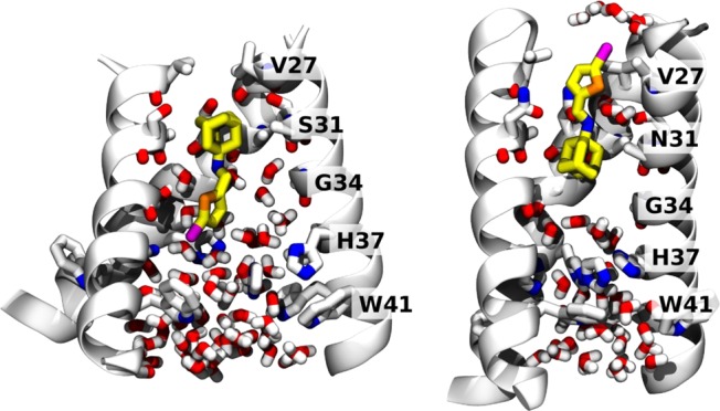
Final MD snapshots of compound 11 bound to the transmembrane segment of WT and S31N M2 in a lipid bilayer. Left: 11 bound within WT M2 after 200 ns. Right: 11 bound within S31N M2 after 100 ns. The protein backbone is shown as ribbons; pore-lining side chains and backbone carbonyls, ligand, and water molecules within the pore are shown as sticks. Water molecules solvating the bromine atom in the outward-facing pose (right) are also shown.