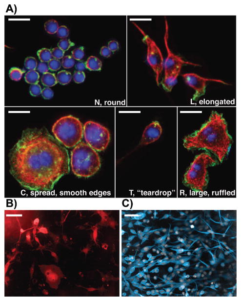
Figure 1.
Morphological complexity in different cell lines. A: The five shapes adopted by wild-type Drosophila Kc Hemocytes [23]. We have termed the shapes “N”, “L”, “C”, “T”, and “R”. Cells were fixed and labeled with Hoechst (blue), phalloidin (green), and anti-tubulin antibody (red). All scale bars represent 20 μm. B: Drosophila BG-2 neuronal cells. BG-2 cells are very heterogeneous, and we have identified six different shapes [29]. BG-2 cells were transfected with EGFP (red) in order to label the entire cell body. Scale bar represents 20 μm. C: WM266.4 melanoma cells cultured on collagen and labeled with CellTracker dye and DAPI. Melanoma cells adopt two types of shape: rounded and elongated. Scale bar represents 50 μm.

