
Figure 2. Loss of KLF5 affects cytodifferentiation.
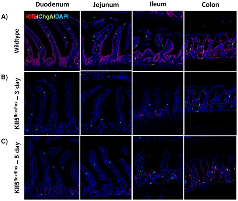
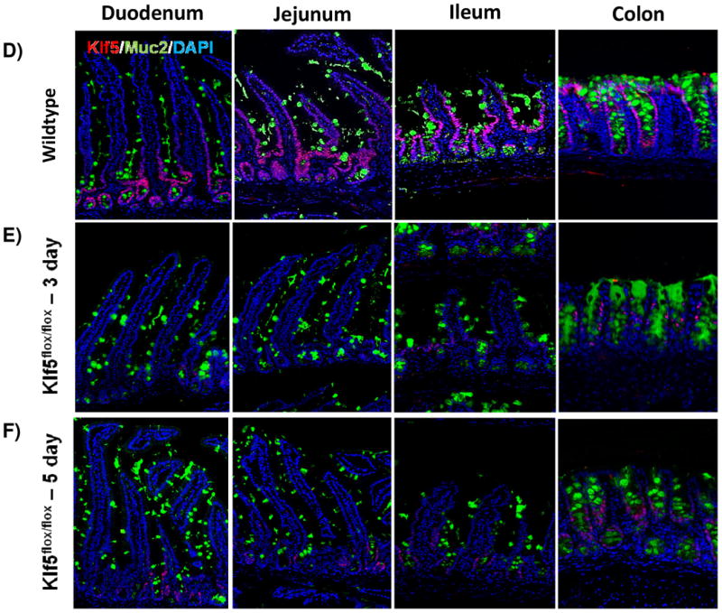
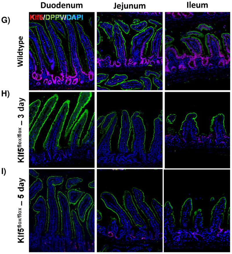
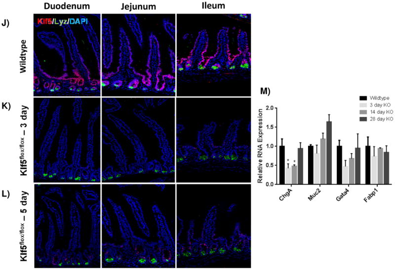
Histological comparison of Klf5loxP/loxP; VilWT (WT) and Klf5loxP/loxP; VilCreERT2 (Klf5-mutant) intestinal tissues for cellular differentiation utilizing immunofluorescent staining for chromagranin A (green; enteroendocrine cells) (A-C),mucin2 (green; goblet cells) (D-F), lysozyme (green; Paneth cells) (G-I), and dipeptidyl peptidase-4 (green; enterocytes) (J-L). Nuclear stain is DAPI (blue). (M) Comparison of ChgA, Muc2, Gata4, and Fabp1 mRNA expression analysis by RT-qPCR from jejunal segments of the small intestine. Student's t-test was performed to determine p-values; expression was normalized to Gapdh. (Mean ± SEM: P ≤ 0.05, **P ≤ 0.01, ***P ≤ 0.001, n = 3-6).