
Figure 3. Loss of KLF5 attenuates cellular proliferation in the adult intestine.
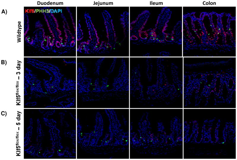
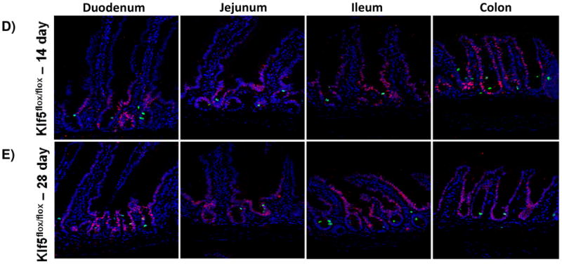
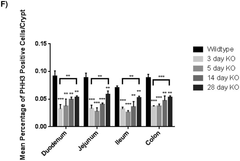
(A) Histological analysis for the comparison of cellular proliferation utilizing immunofluorescent staining for mitotic marker, phosphohistoneH3 (green) in wildtype and Klf5 loss-of-function mice (3, 5, 14, and 28 days post tamoxifen-induced recombination). Nuclear stain is DAPI (blue). (B) Quantification of average percentage of phosphohistoneH3 positive cells per crypt. Significance determined between groups of two (wildtype vs. loss of Klf5 for 3 days, wildtype vs. loss of Klf5 for 5 days, wildtype vs. loss of Klf5 for 14 days, wildtype vs. loss of Klf5 for 28 days, and loss of Klf5 for 3 vs. 28 days). (Mean ± SD, T-test: * P ≤ 0.05, **P ≤ 0.01, ***P ≤ 0.001, n = 3)