
Figure 4. Intestinal crypt cell death is increased early after loss of KLF5, but disappears over time.
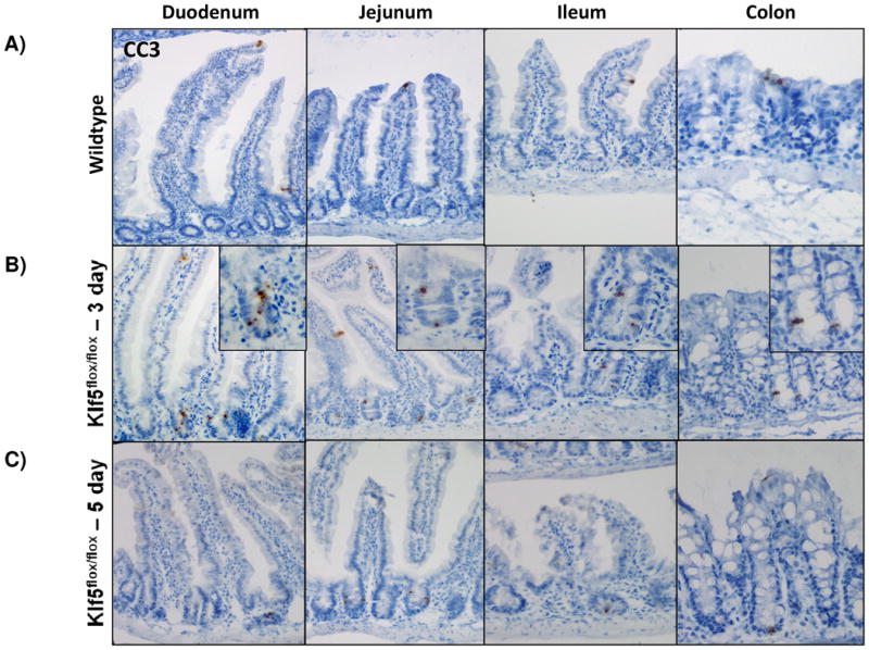
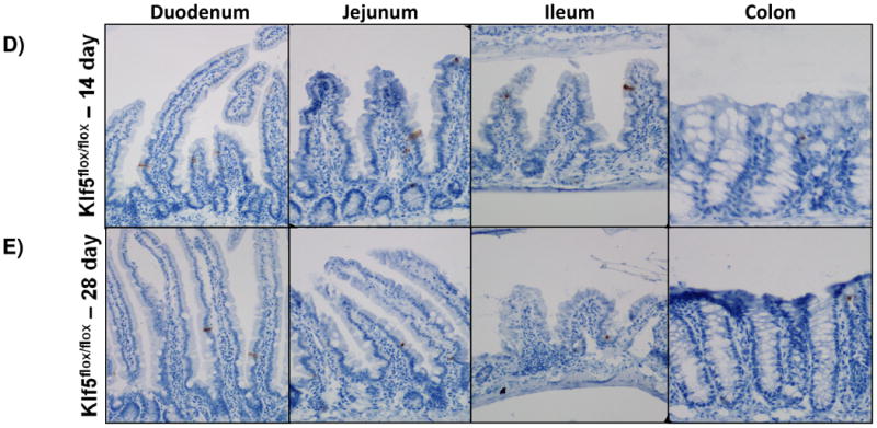
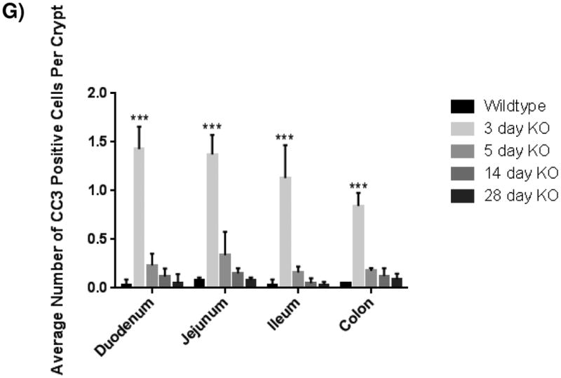
(A-E) Immunohistochemical analysis of apoposis is shown using cleaved-caspase 3 (brown) counterstained with hemotoxylin (blue) in wildtype and Klf5 loss-of-function mice (3, 5, 14, 28 days post tamoxifen-induced recombination). (F) Quantification of the number of cleaved-caspase 3 positive cells per crypt. Significance determined between groups of two (wildtype vs. loss of Klf5 for 3 days, wildtype vs. loss of Klf5 for 5 days, wildtype vs. loss of Klf5 for 14 days, wildtype vs. loss of Klf5 for 28 days, and loss of Klf5 for 3 vs. 28 days). (Mean ± SD, T-test: * P ≤ 0.05, **P ≤ 0.01, ***P ≤ 0.001, n = 3)