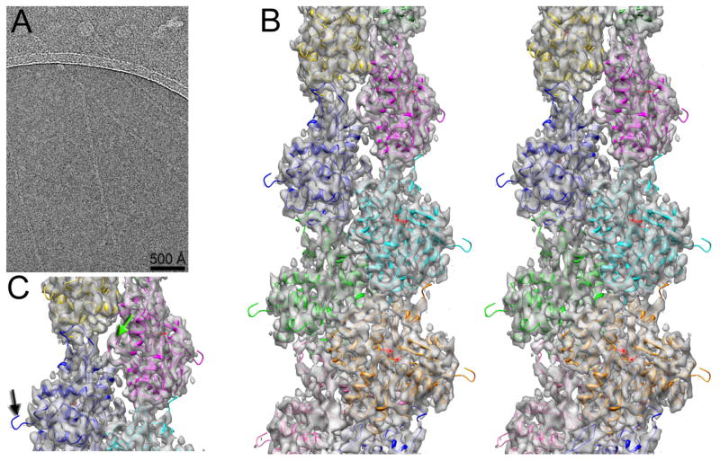
Fig. 1.
3D-reconstruction of frozen hydrated actin filaments. (A) Typical micrograph of actin filaments embedded in thin ice. (B) Stereo view of the 3D-reconstruction at ~ 4.7 Å resolution. (C) The absence of the N-terminal density in the map is indicated with a black arrow, while the hydrophobic plug density is marked with a green arrow.

