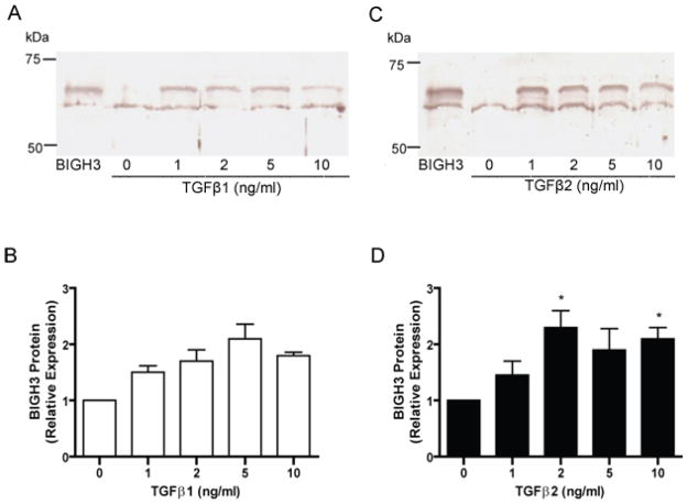
Figure 4. TGFβ 1 and 2 Induced BIGH3 Protein Secretion by RhREC.
RhRECs were cultured with either TGFβ1 (A) or TGFβ2 (B) at 1, 2, 5 and 10 ng/mL for 24 hrs and refreshed media were collected after a 24 hrs period and probed for the BIGH3 protein by Western blot. Lane 1, 0.1 μg recombinant BIGH3 protein; lane 2–6, BIGH3 protein in cell media of cells treated with TGFβ at 0 ng/mL, 1 ng/mL, 2 ng/mL, 5 ng/mL, 10 ng/mL. Densitometry results from A and C respectively are presented in (B) and (D). One way ANOVA showed no significant dose effect for TGFβ1 but a significant dose effect for TGFβ2 [F(4,14)= 4.081; P<0.05]. Dunnett’s test (D) was performed using 0 ng/mL as control (*P<0.05).

