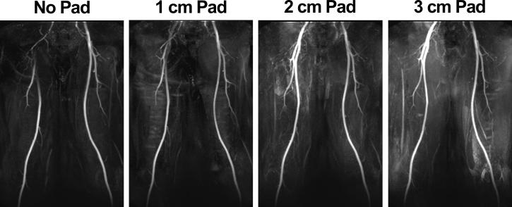
Figure 1.
MIPs of a volunteer with three different high-permittivity pad thicknesses: 1 cm (second column), 2 cm (third column), and 3 cm (fourth column). For reference, a MIP without padding is also shown (first column). All four MIPs were displayed in identical grayscales. Pad placement was on the upper-thigh to the pelvis following the path of the femoral arteries. Based on this preliminary analysis, we used the 2 cm thick pad throughout this study.

