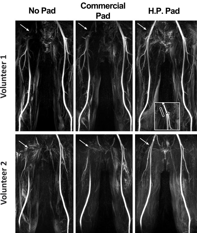
Figure 3.
Representative MIPs of from two volunteers, volunteer 1 on top and volunteer 2 on bottom, with three different settings: baseline (left), commercial dielectric padding (middle), and high-permittivity dielectric padding (right). Compared with baseline and commercial dielectric padding, high-permittivity dielectric padding significantly increased signal around the bifurcation point of the common femoral artery. MIPs displayed in identical grayscales per subject.

