Figure 2.
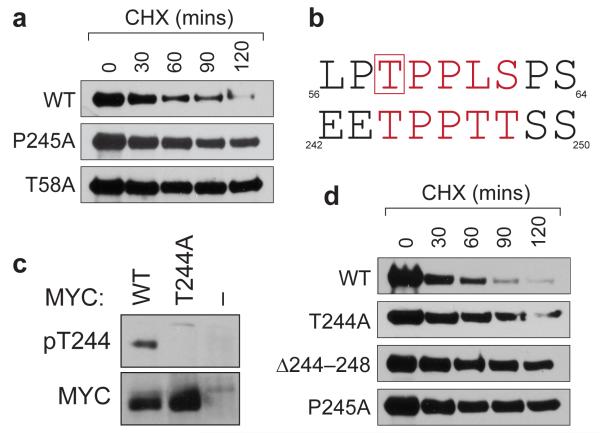
The 244–248 segment of MYC is a phosphodegron that is inactivated by the P245A tumor mutation. (a) Site-directed mutagenesis was used to introduce the indicated mutations into the MSCV–MYC–IRES–GFP vector, 9 which expresses HA-epitope tagged MYC along with an IRES-driven green fluorescent protein (GFP). These constructs were retrovirally transduced into NIH3T3 cells. To infer MYC stability, stable transductants were treated with cycloheximide (CHX) for the indicated times to inhibit protein synthesis, 22 protein extracts prepared, and exogenous MYC levels determined by Western blotting with an anti-HA antibody. (b) Alignment of MYC residues 56–64 and 242–250. Core residues of the SCFFbw7 phosphodegron are in red. Residue T58, which must be phosphorylated to bind SCFFbw7, is boxed. (c) NIH3T3 cells expressing the indicated HA-tagged MYC proteins (or vector control ‘–’) were lysed, HA-tagged proteins recovered by immunoprecipitation, and either T244-phosphorylated, or total, MYC proteins detected by Western blotting. Characterization of the specificity of purified phospho-T244 antibodies is presented in Supplementary Figure S2. (d) As in (a) except that NIH3T3 cells expressed WT, T244A, Δ244–248, or P245A forms of MYC.

