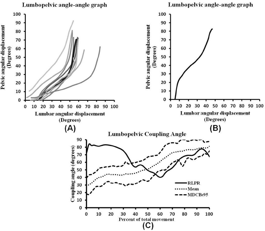
Figure 3.
Angle-angle and coupling angle graphs of forward bend derived from individuals with typical and a single subject with reversed lumbopelvic rhythm. (A) Typical lumbopelvic rhythm angle-angle graphs from 10 individuals without low back pain and clinically observed typical lumbopelvic rhythm. (B) Reversal of lumbopelvic rhythm angle-angle graph during forward bending phase in an individual with low back pain and clinically observed reversed lumbopelvic rhythm. (C) Coupling angle-movement graph where the kinematic data of an individual with observed reversal of lumbopelvic rhythm (solid line) were plotted on a graph representing a typical profile (dotted line) with 95% minimal detectable change bands (dashed lines) created from 15 healthy subjects visually rated as having a typical pattern of forward bending motion. The first 35% of the movement demonstrates a radically altered pattern from typical coordination of the lumbar spine and pelvis during this task.

