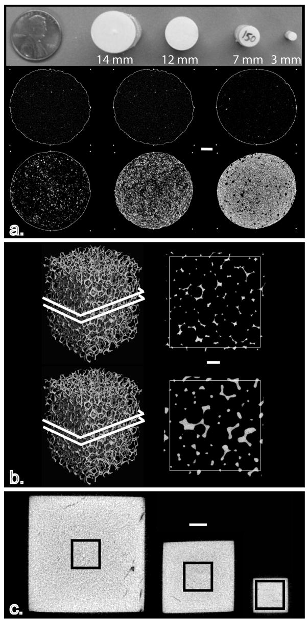
Figure 1.

Image showing all the models used to assess the influence of size on the measurement of ρm using μCT: (a) hydroxyapatite rods ranging in size from 3–14 mm and varying in hydroxyapatite concentration, (b) precision-made porous aluminum foams ranging in aluminum volume fraction from approximately 5% to 12% and, (c) bovine cortical bone cubes were extracted and then alternately scanned and reduced in size twice. White scale bars = 2 mm
