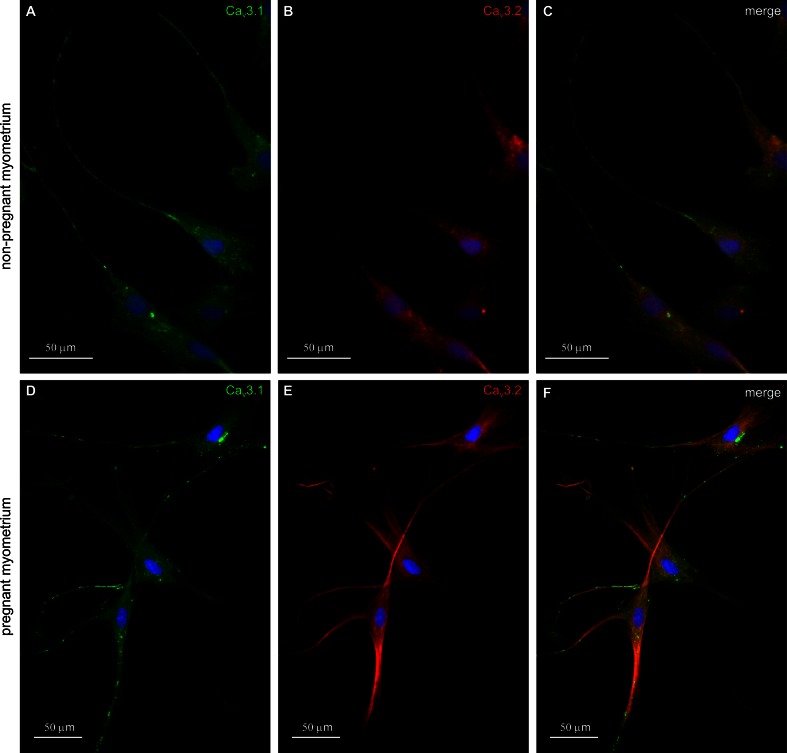
Fig. 2.
Double immunolabeling for T-type calcium channels in cell cultures from non-pregnant/pregnant myometrium. a CaV3.1 immunolabeling (green) was detected on TCs cell body, but it was stronger at Tps level, b CaV3.2 expression was detected only at cell body level. c Co-expression of CaV3.1-positive (green) and CaV3.2 (red) is presented on merged images. d Immunolabeling for T-type calcium channels in cell cultures from pregnant myometrium. Strong staining for CaV3.1 was found in the cytoplasm, adjacent to the nucleus and in the Tps of the TCs. e CaV3.2 immunolabeling was found throughout the cytoplasm of TCs and predominantly within Tps. f Merged images show co-expression pattern for CaV3.1 (green) and CaV3.2 (red). Note that both TCs and SMCs (cells with widened cell body without extensions) express CaV3.1 and CaV3.2. Nuclei were stained with DAPI. Bar 50 µm

