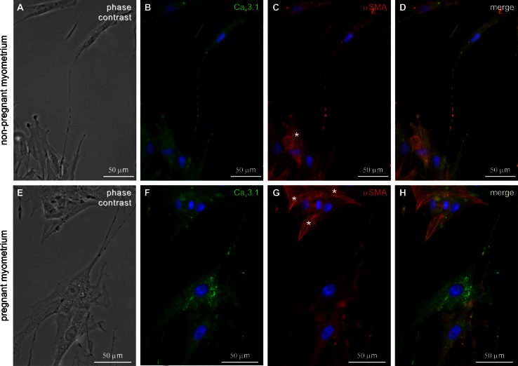
Fig. 4.
Double immunolabeling for CaV3.1 and α-SMA in cell cultures from non-pregnant/pregnant myometrium. a, e Phase contrast. b, f Strong staining for CaV3.1 was found on both cell body and Tps of the TCs. c, g αSMA immunolabeling was found in SMCs [cells with widened bodies and without cellular extensions (asterisks)]. d, h Merged images of CaV3.1 and α-SMA. Nuclei were stained with DAPI. Bar 50 µm

